28621583
Description
Flashcards by Katrina Oschmann, updated more than 1 year ago
|
|
Created by Katrina Oschmann
over 4 years ago
|
|
| Question | Answer |
| genome | all of a cell's DNA |
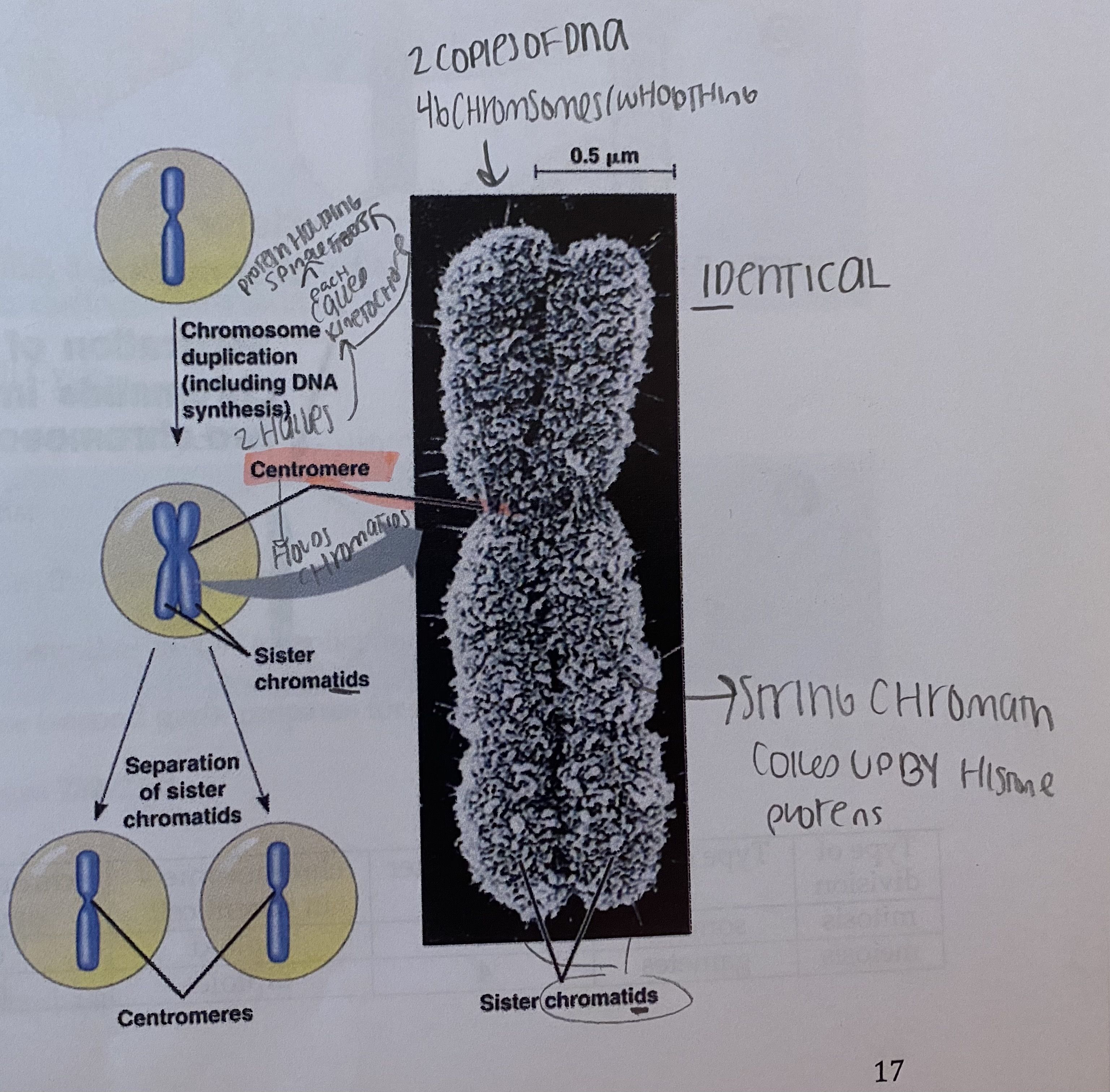
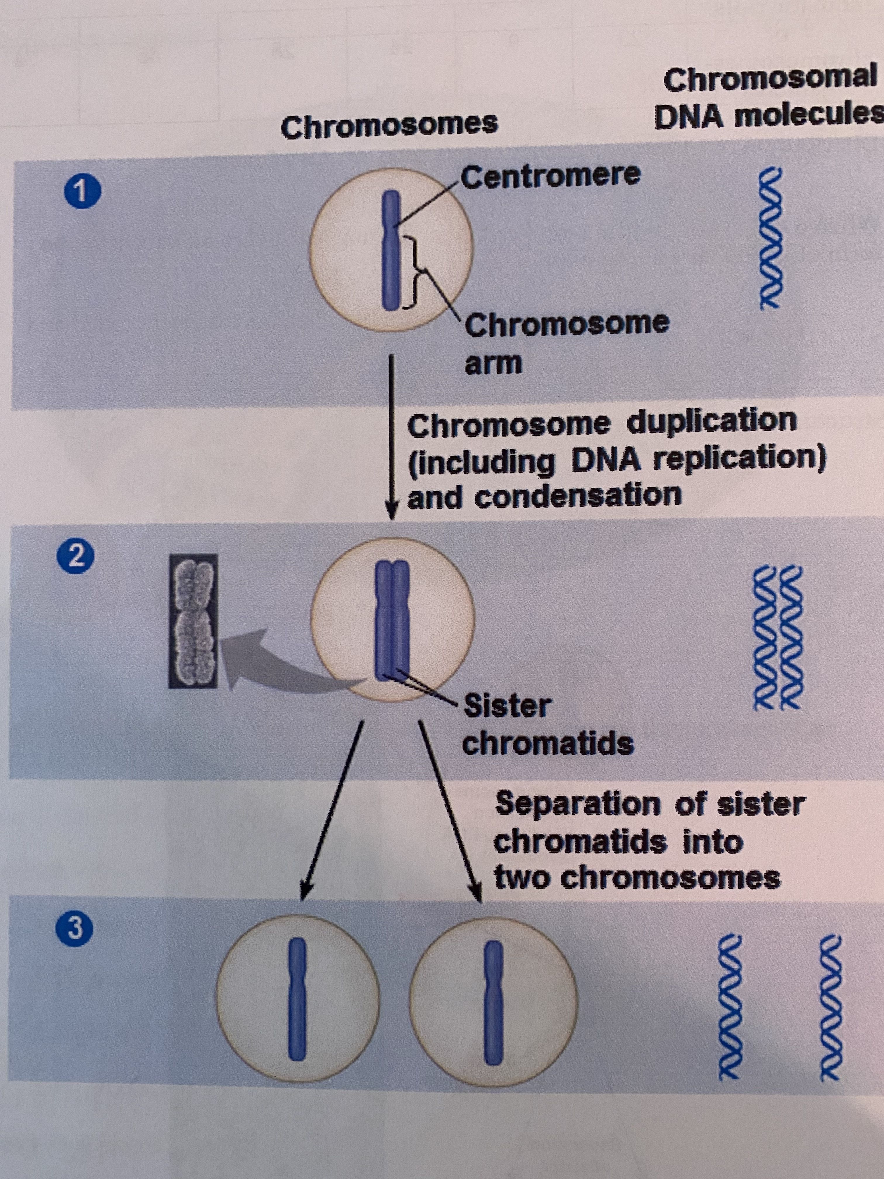
| chromosome - structure/parts the name for the package holding one very long DNA molecule in a cell; carries several hundred to thousands of genes, the DNA is coiled up by histone proteins - see other cards about duplication, structure, etc. | |
| histone proteins | a protein involved in DNA coiling |
| chromatin | uncoiled chromosomal material (interphase) - the form of each chromosome when it is not dividing and is replicated is a long chromatin fiber - once they replicate (chromosomes), each chromatin fiber is densely coiled and folded |
| somatic cells | all body cells, are diploid |
| gametes | reproductive cells - sperm and eggs; are haploid |
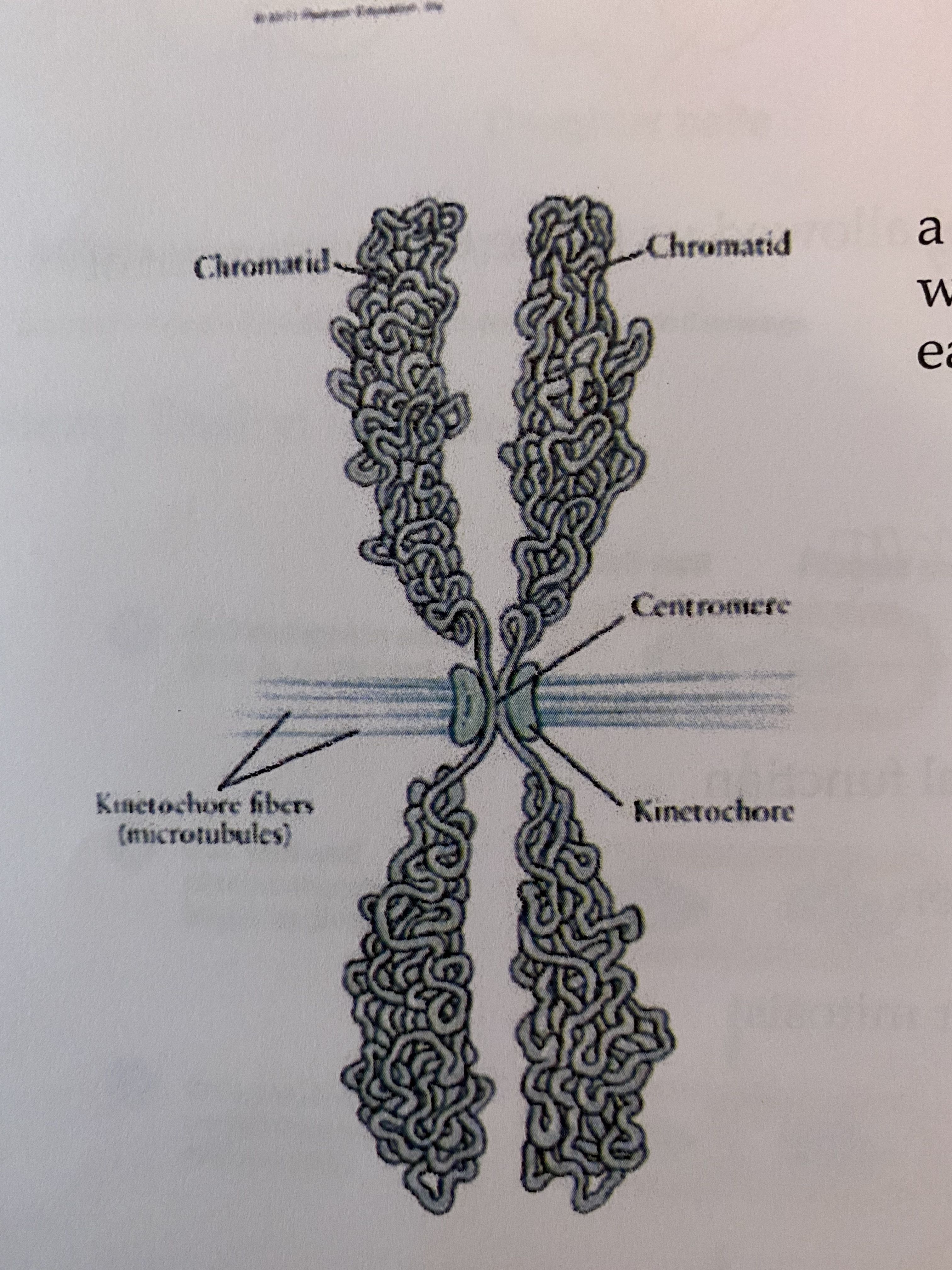
| sister chromatids Replicated forms of a chromosome joined together by the centromere and eventually separated during mitosis or meiosis II. identical | |
| centromere Area where the chromatids of a chromosome are attached | |
| diploid | (of a cell or nucleus) containing two complete sets of chromosomes, one from each parent. |
| haploid | (of a cell or nucleus) having a single set of unpaired chromosomes. |
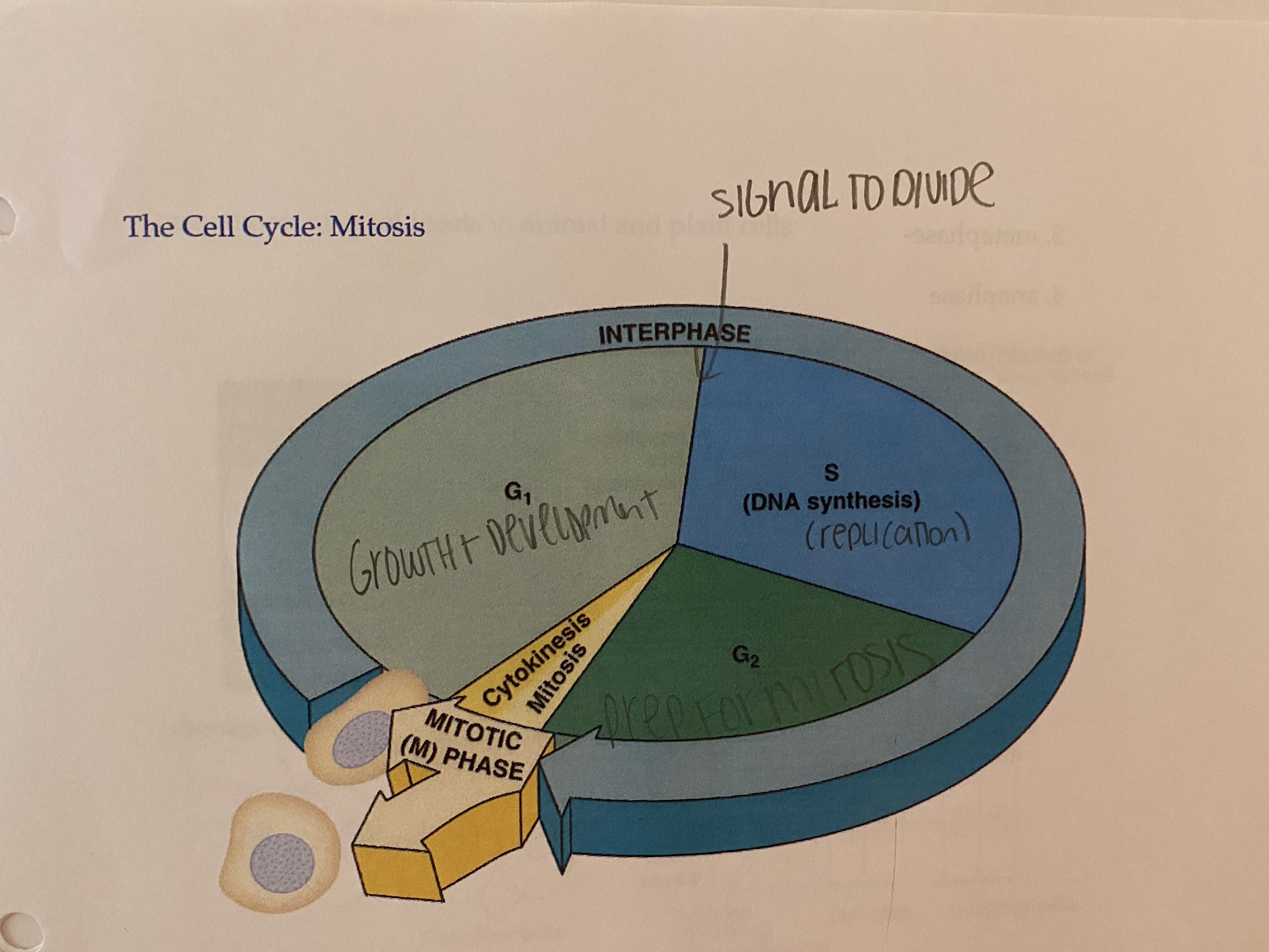
| cell cycle (diagram) A) interphase (90%) - G1 phase (first gap): growth, normal function - S phase (synthesis): DNA replication - G2 phase (second gap): prepares for mitosis B) mitosis - prophase - prometaphase - metaphase - anaphase - telophase C) cytokinesis (part of mitotic (M) phase) | |
| Walter Fleming | developed the stain that allowed us to see the movement of the chromosomes during cell division |
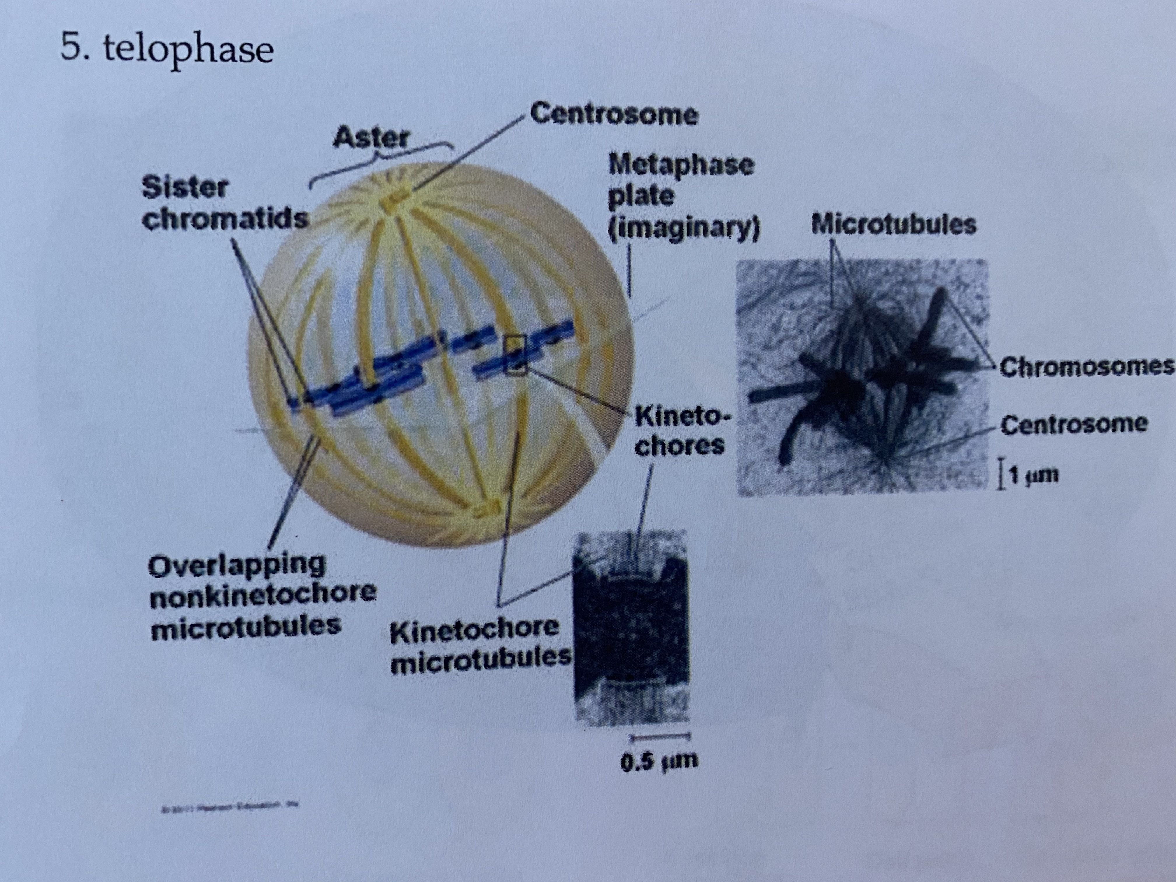
| kinetochore | structure of proteins associated with specific sections of chromosomal DNA at each centromere - 2 face in opposite direction |
| kinetochore microtubules | during prometaphase, some of the spindle microtubules attach to the kinetochores so they are called kinetochore microtubules - come from opposite poles, they tug on the chromosome until it is positioned on the equator at metaphase |
| nonkinetochore microtubules | responsible for elongating the whole cell during anaphase |
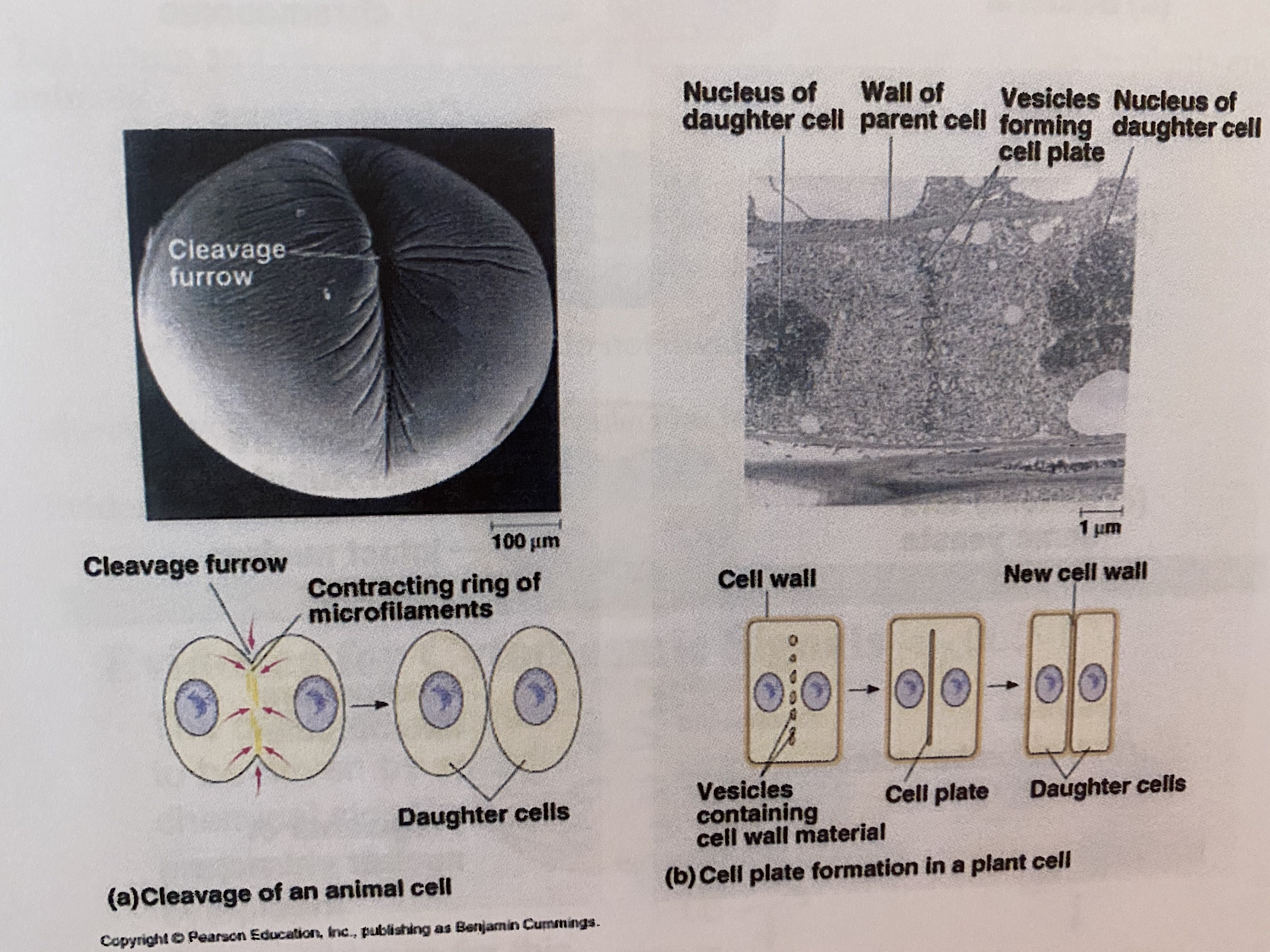
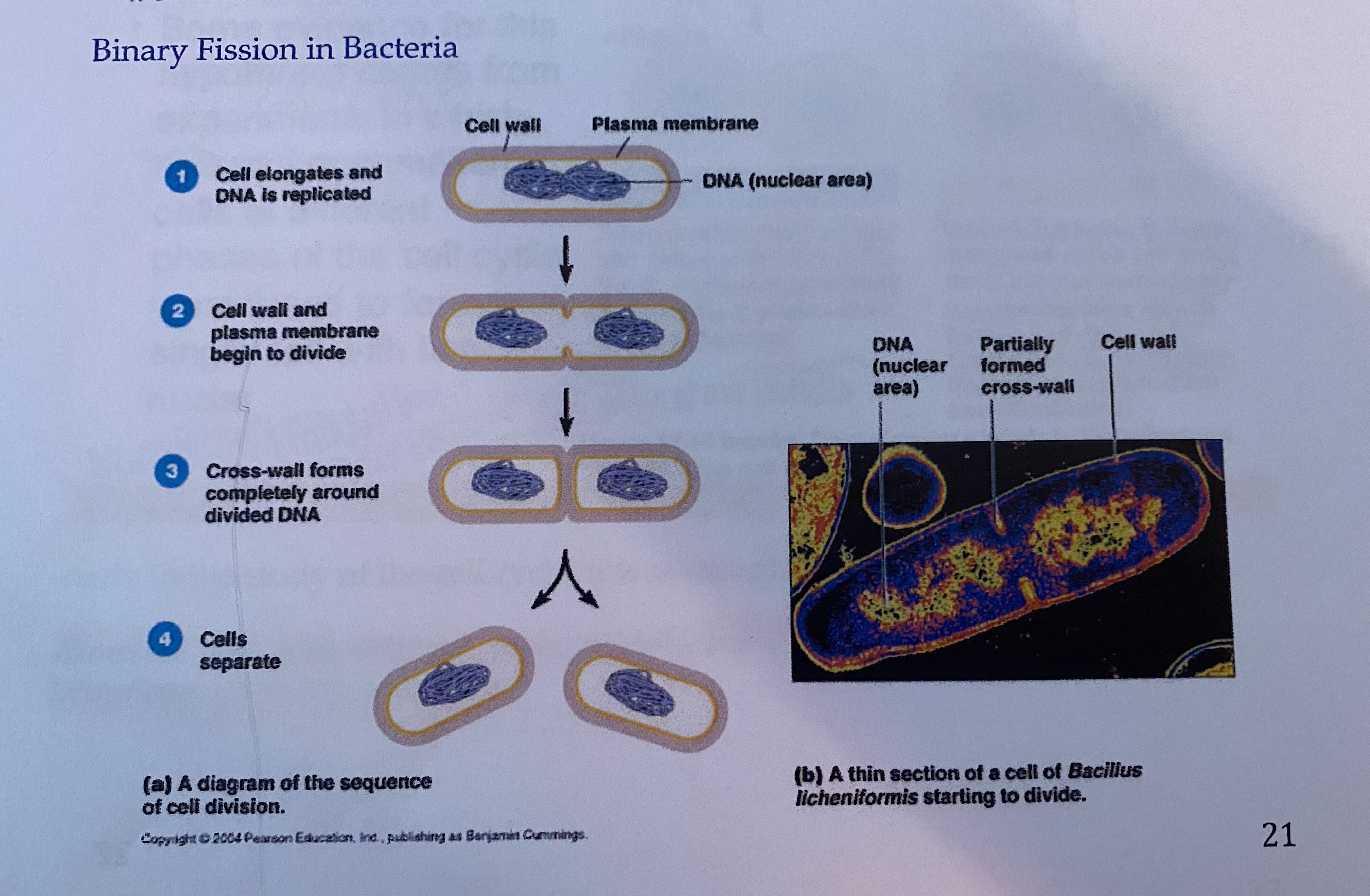
| How does cytokinesis differ in plant and animal cells? in animal cells - cleavage furrow containing ring of microfilaments > daughter cells plant cells - cell wall > vesicles containing cell wall material material . cell plate > daughter cell | |
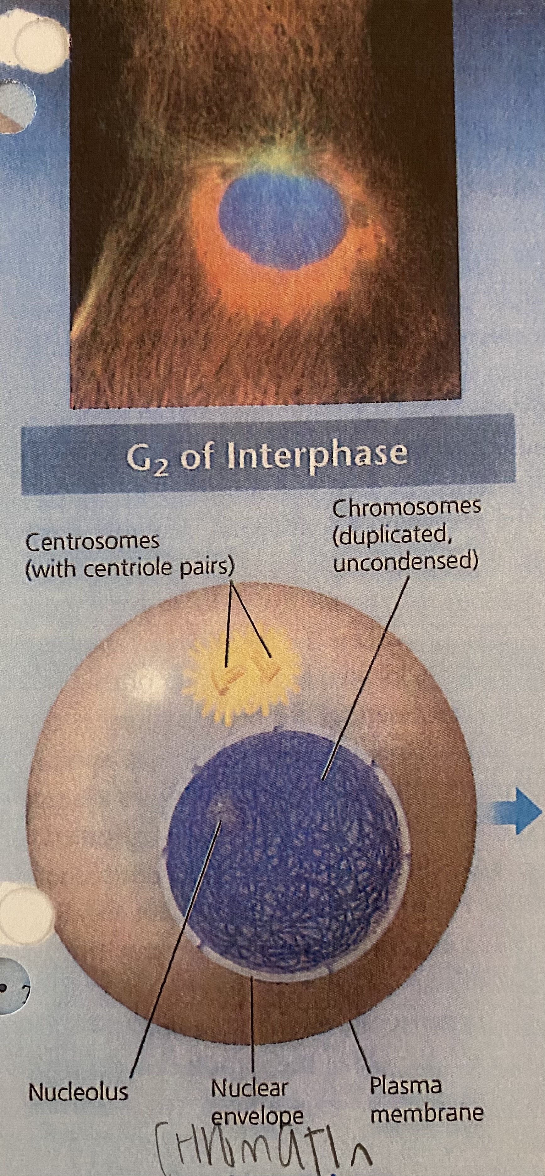
| G2 Interphase * a nuclear envelope encloses the nucleus *the nucleus contains one or more nucleoli (nucleolus) * two centrosomes have formed by duplication of a single centrosome. centrosomes are regions in animal cells that organize the microtubules of the spindle. each centrosome contains two centrioles. *chromosomes, duplicated during S phase cannot be seen individually because they have not been condensed yet centrosomes - microtubule organizing center (animal cells) plant cells form a mitotic spindle without centrioles | |
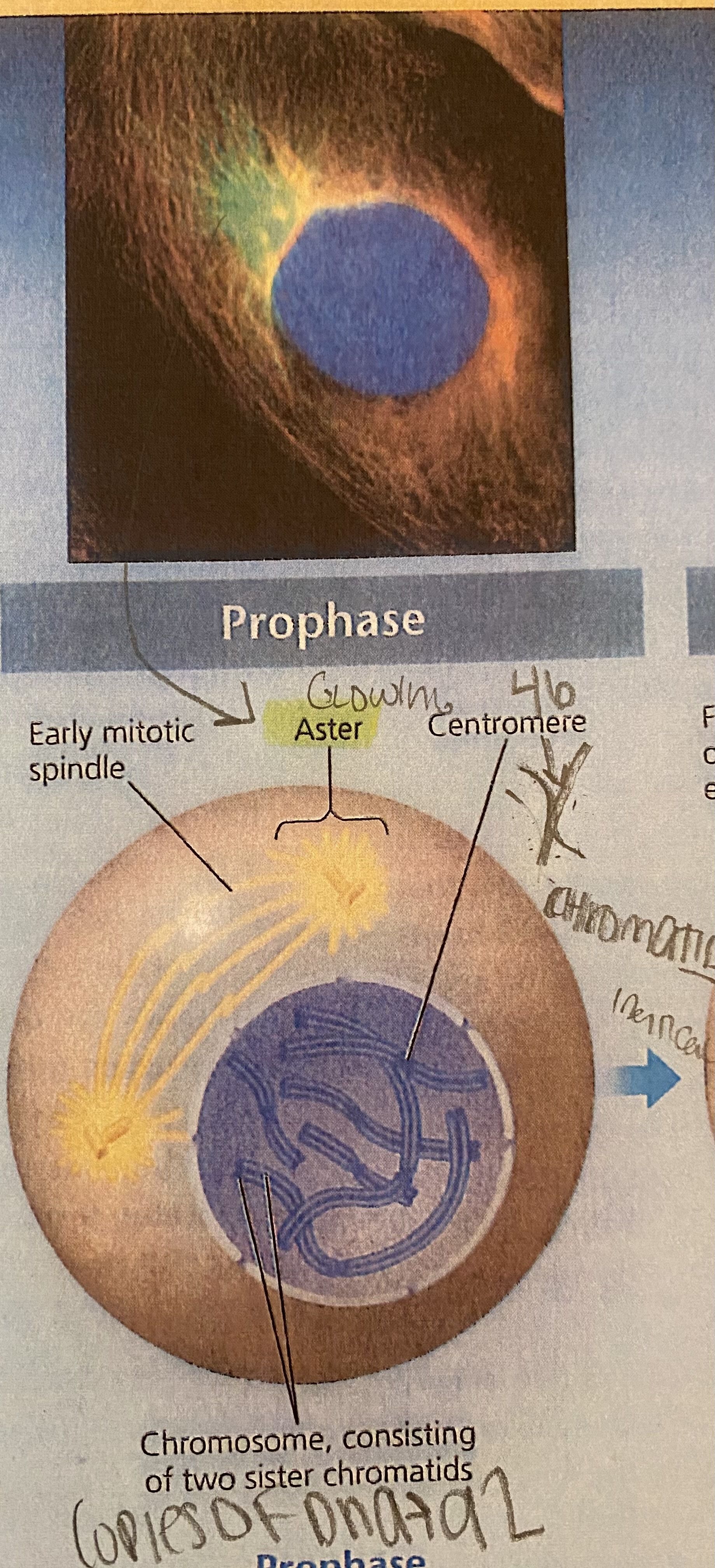
| prophase *the chromatin fibers become more tightly coiled, condensing into discrete chromosomes observable with a light microscope * the nucleoli disappear * each duplicated chromosomes appears as two identical sister chromatids joined at their centrosomes and, in some species, all along their arms by cohesions (proteins that hold chromatids together...called sister chromatid cohesion) * the mitotic spindle (named for its shape) begins to form. it is composed of the centrosomes and the microtubules that extend from them. the radial arrays of shorter microtubules that extend from the centrosomes are called asters ("stars," set of microtubules that extend from centrosome) * the centrosomes move away from each other, propelled partly by the lengthening microtubules between them | |
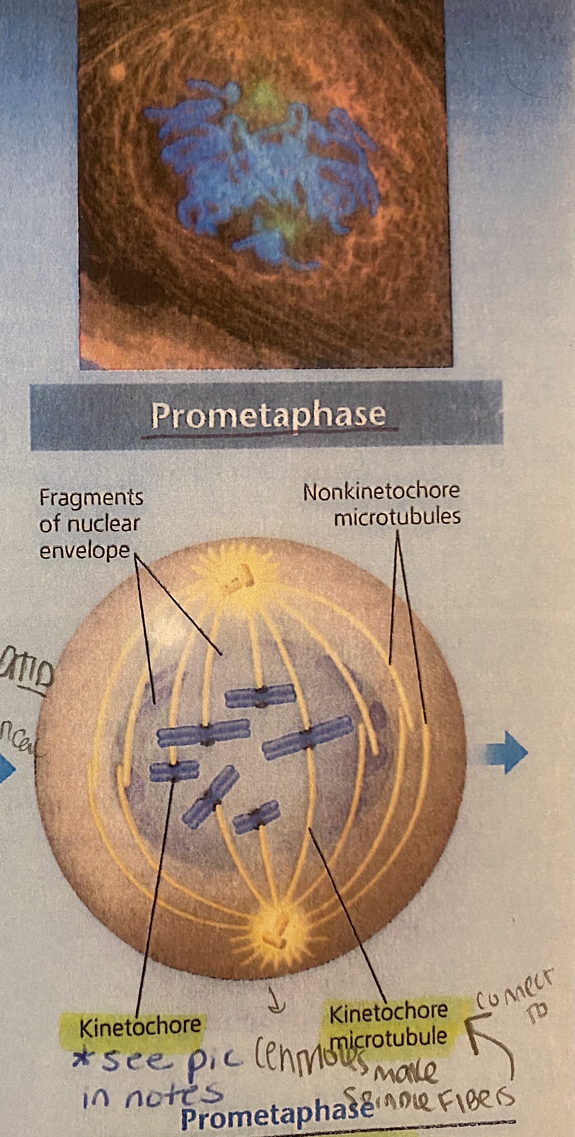
| prometaphase *nuclear envelope fragments (break down) * the microtubules extending from each centrosome can now invade the nuclear area * the chromosomes have become even more condensed * each of the two chromatids of each chromosome now has a kinetochore, a specialized protein structure at the centromere * some of the microtubules attach to the kinetochores, becoming "kinetochore microtubules," which jerk the chromosomes back and forth * nonkinetochore microtubules interact with those from the opposite pole of the spindle | |
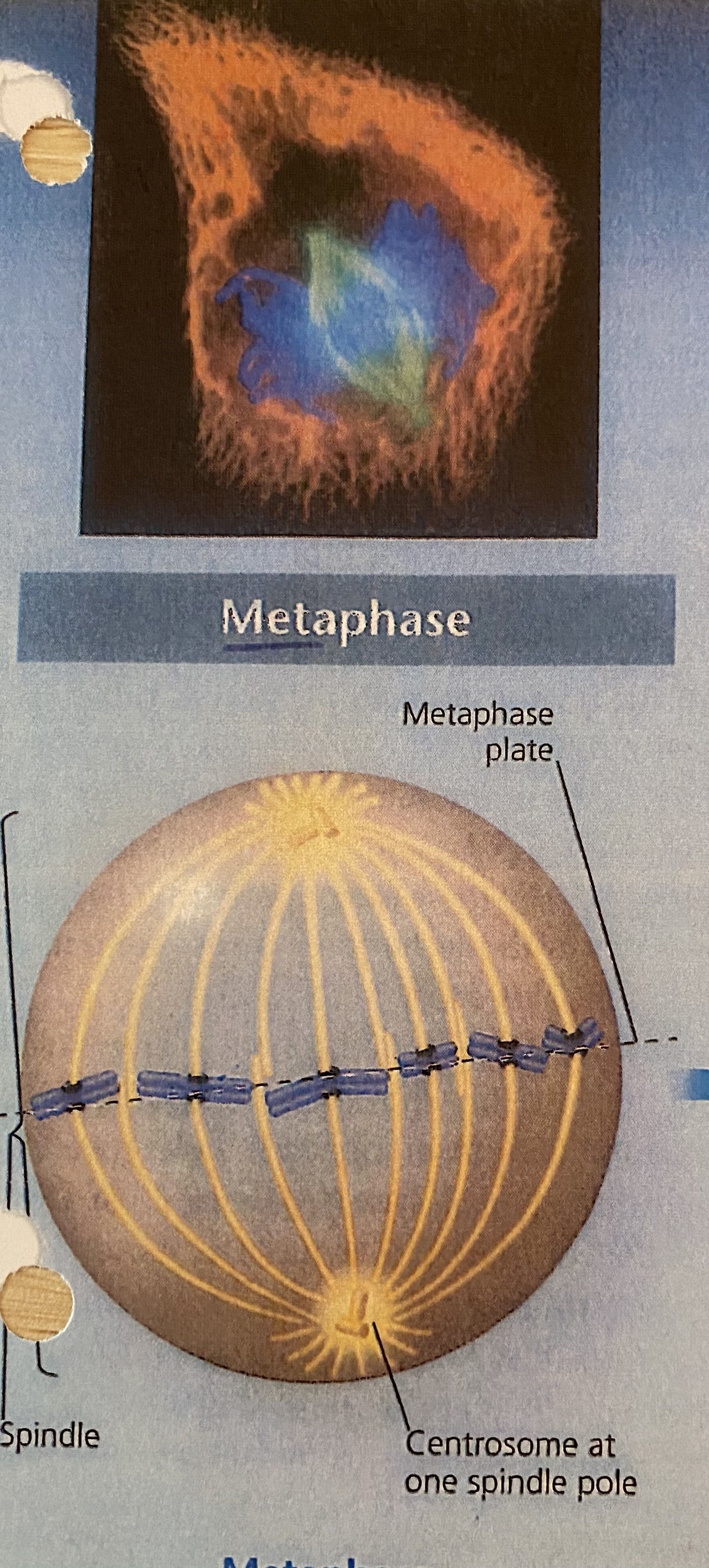
| metaphase *the centrosomes are now at opposite poles of the cell *the chromosomes convene at the metaphase plate, a plane that is equidistant between the spindles two pole. the chromosomes' centromeres lie at metaphase plate. *for each chromosome, the kinetochores of the sister chromatids are attached to kinetochore microtubules coming from opposite poles | |
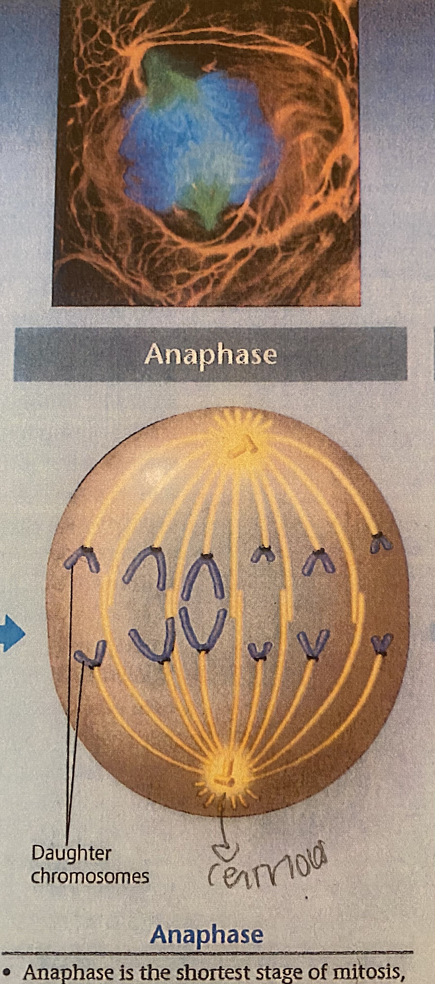
| anaphase *shortest stage of mitosis (only a few minutes) *begins when the cohesin proteins are cleaved (by separase), allowing two sister chromatids of each pair to part suddenly. each chromatid thus becomes a full-fledged chromosome *the two liberated daughter chromosomes begin moving toward opposite ends of the cell as their kinetochore microtubules shorten. because these microtubules are attached at the centromere region, the chromosomes move centromere first *cell elongates as the nonkinetochore microtubules lengthen *by the end of anaphase, the two ends of the cell have equivalent-and complete- collections of chromosomes | |
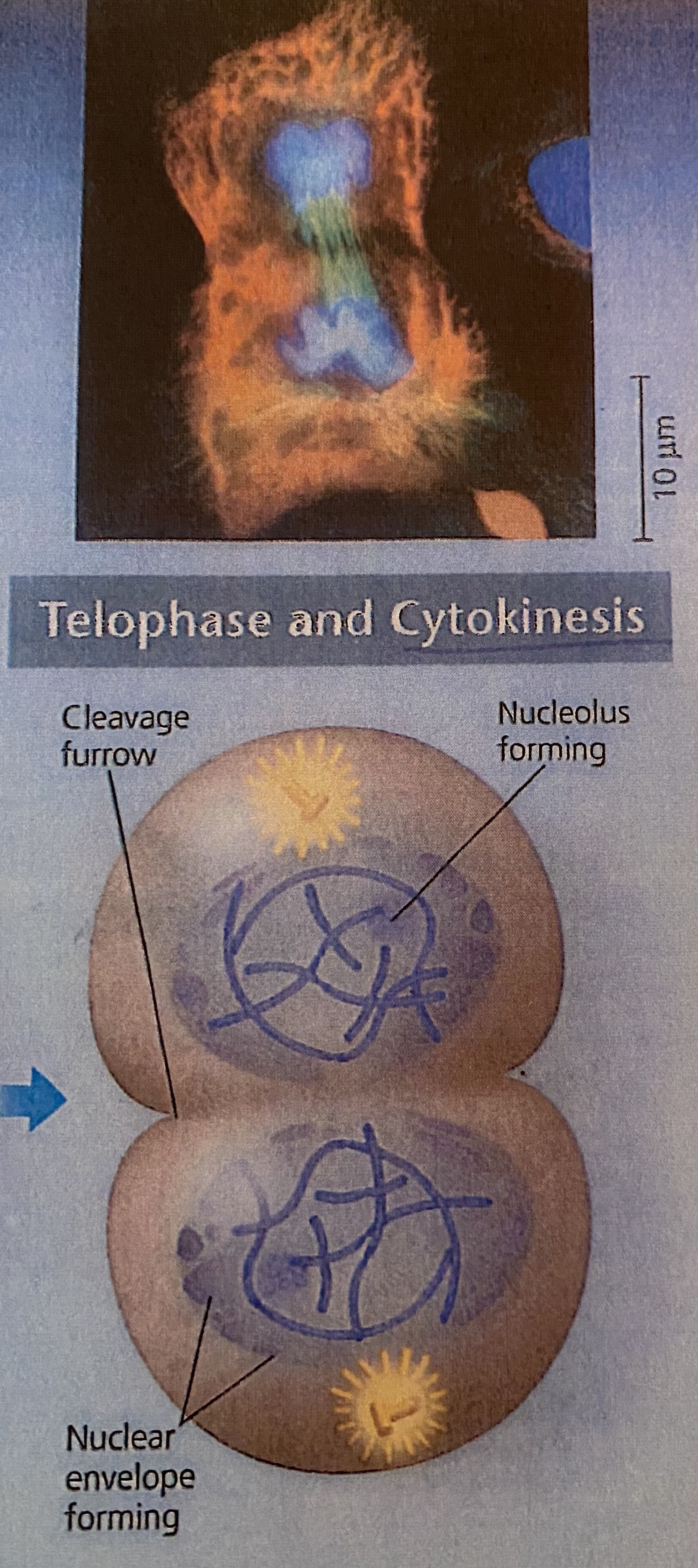
| telophase *the two daughter nuclei form in the cell, nuclear envelopes arise from the fragments of the parent cell's nuclear envelope and other portions of the endomembrane system *nucleoli reappear *the chromosomes become less condensed *any remaining spindle microtubules are depolymerized *mitosis, the division of one nucleus into two genetically identical nuclei, is now complete | |
| cytokinesis | *the division of the cytoplasm is usually well under way by late telophase, so the two daughter cells appear shortly after the end of mitosis *in animal cells, cytokinesis involves the formation of a cleavage furrow, which pinches the cell in two |
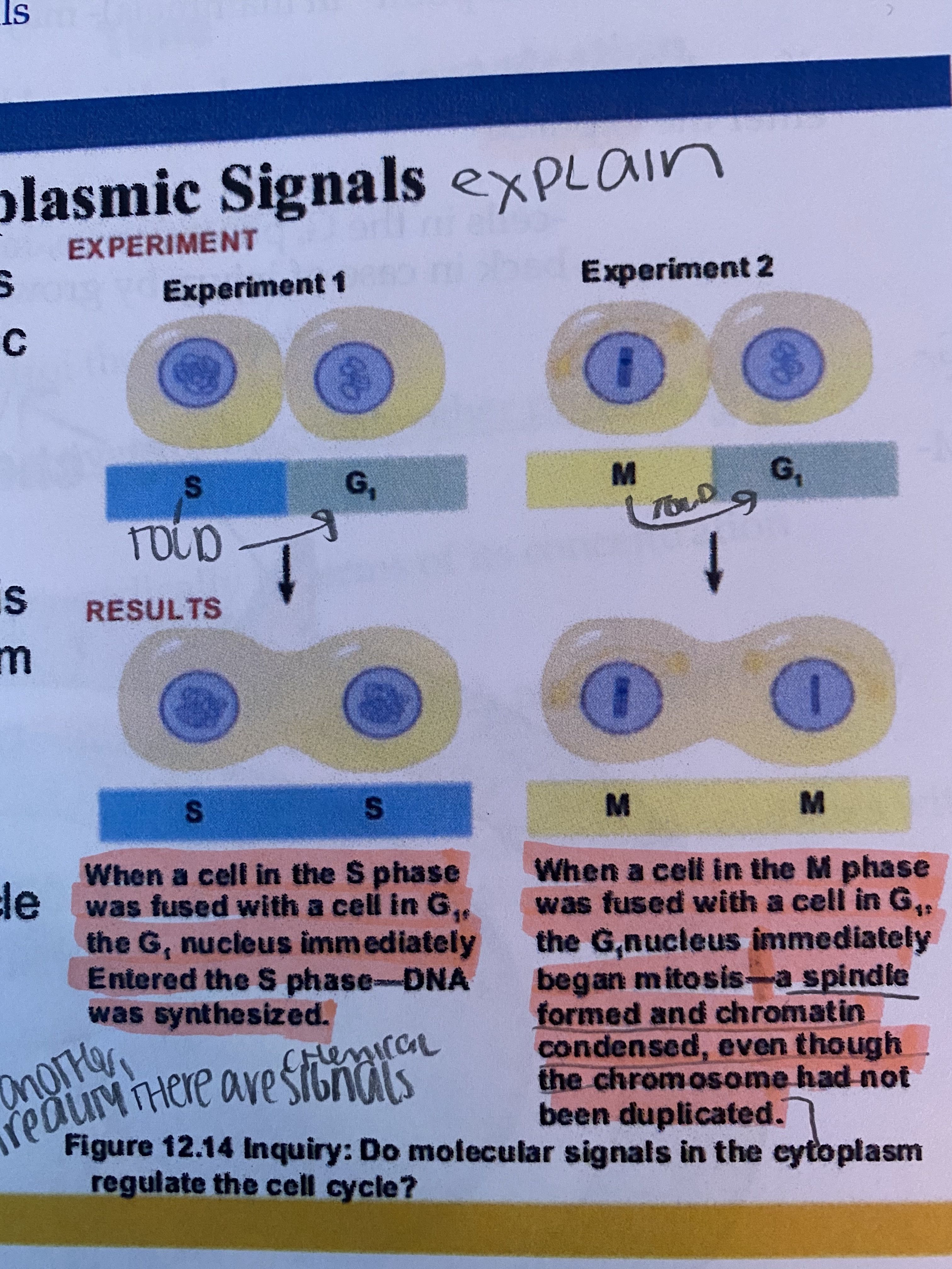
| evidence for cytoplasmic signals experiments the cell cycle appears to be driven by specific chemical signals present in the cytoplasm *some evidence for this hypothesis comes from experiments in which cultured mammalian cells at different phases of the cell cycle were fused to form a single cell with two nuclei experiment 1: when a cell in the S phase was fused with a cell in G1, the G1 nucleus immediately entered the S phase - DNA was synthesized experiment 2: when a cell in the M phase was fused with a cell in G1, the G1 nucleus immediately began mitosis - a spindle formed and chromatin condensed, even though the chromosome has not been duplicated | |
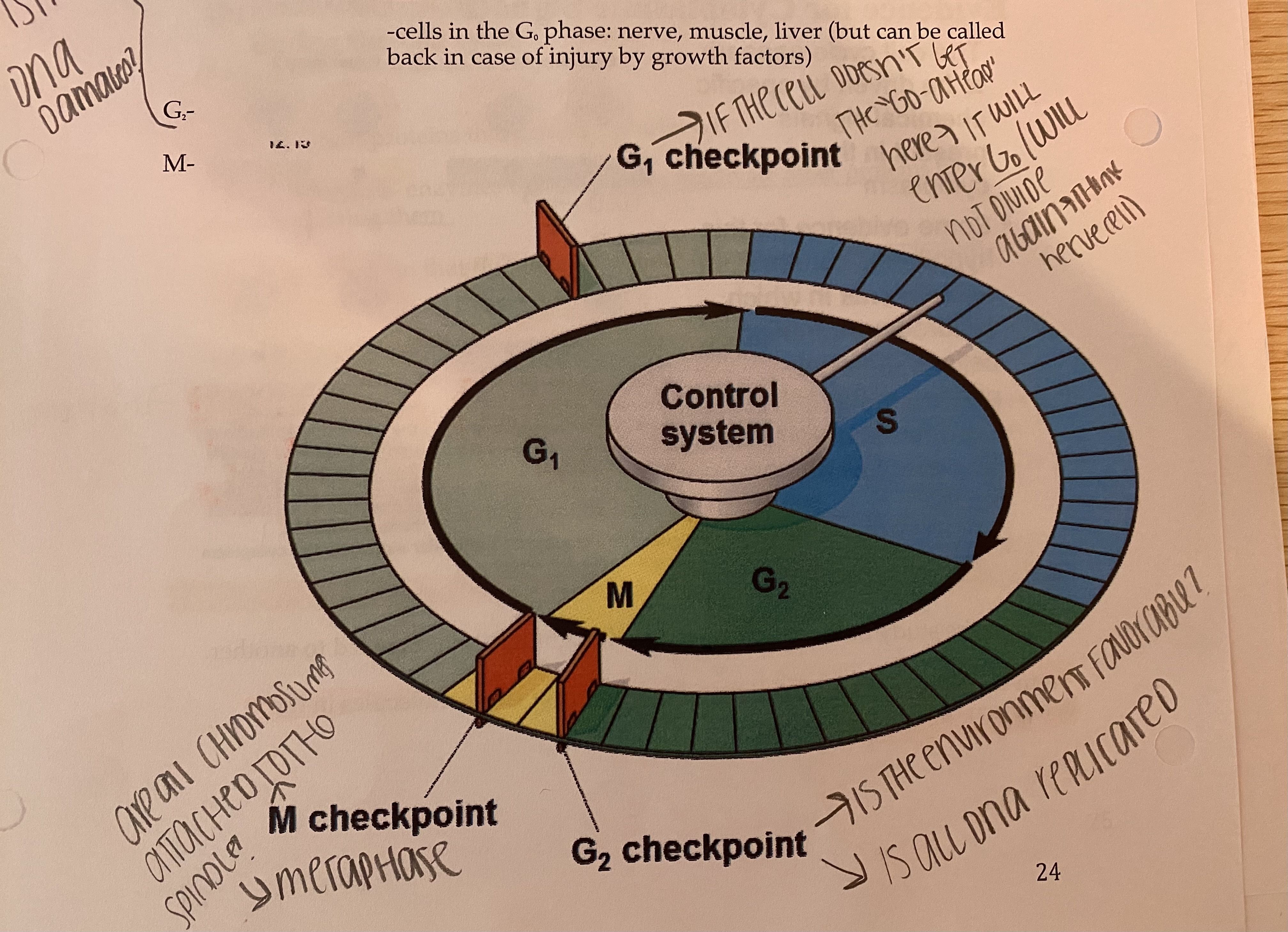
| 3 major cell checkpoints is the DNA damaged? G1 - aka the "restriction point" in mammals (may be most important), if the cell doesn't receive the go-ahead signal here, it will exit the cell cycle and enter G0 phase and never divide again (nerve, muscle, and liver phase in G0 unless called back in case of injury by growth factors) is the DNA damaged (S phase follows and you don't want damaged DNA replicating) does cell have the resources it needs? G2 - is the environment favorable? is all DNA replicated correctly? is it growing well? does it have the resources it needs to continue? M - are all chromosomes attached to the spindle in the middle? (metaphase) (if not, chromosomes won't be separated correctly) *if cell doesn't pass a cell checkpoint because of an issue, apoptosis should occur (sometimes they will be fixed, or cancer) | |
| why will cell enter G0? | the cell doesn't receive the go-ahead signal at the G1 checkpoint, it will exit the cell cycle and enter G0 phase and never divide again (nerve, muscle, and liver phase in G0 unless called back in case of injury by growth factors) |
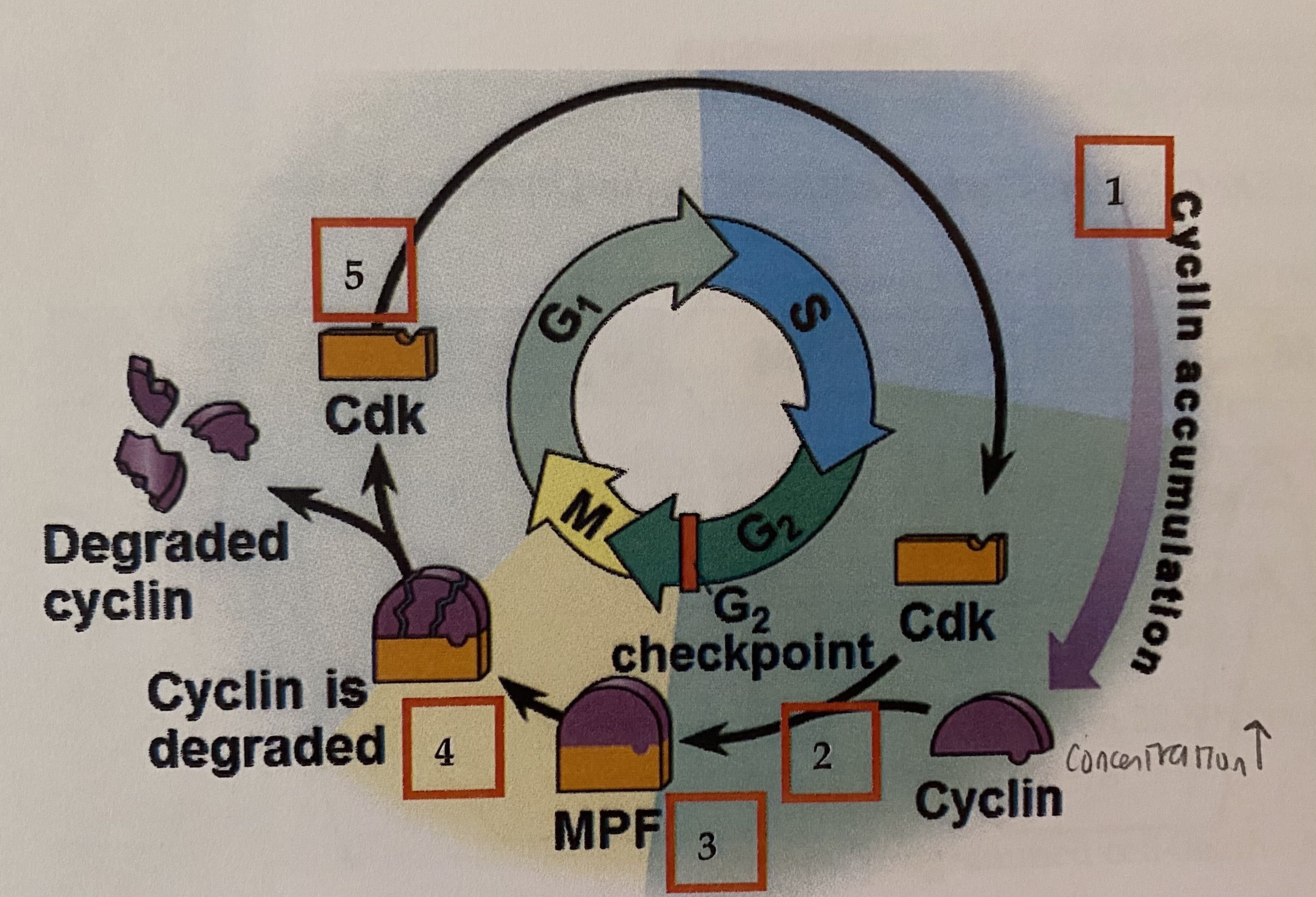
| kinases 1) protein kinase - enzymes that activate other proteins by phosphorylating them 2) cyclins - a protein that fluctuates cyclically in terms of its concentration - some kinases are cyclin-dependent kinases (Cdks) which means that they must be attached to a cyclin in order to work | |
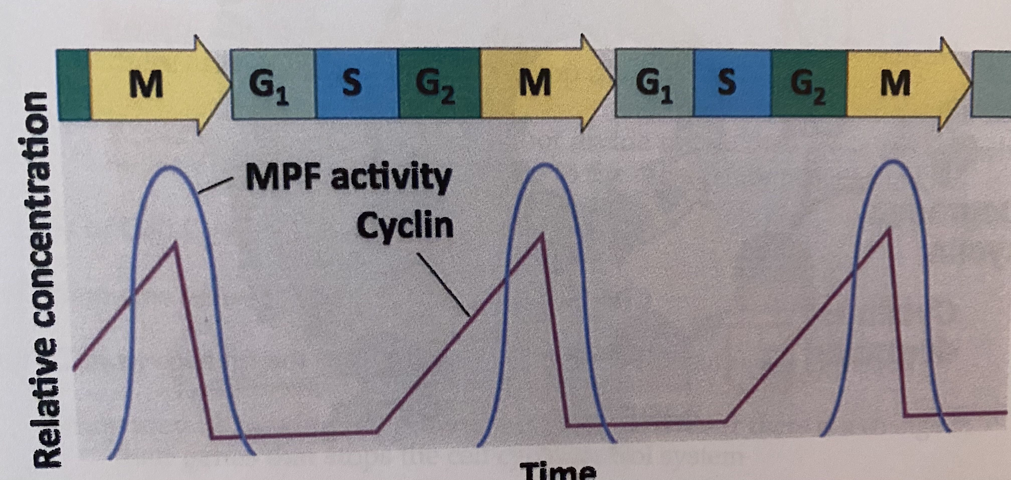
| MPF (and the long list of steps) MPF (maturation promoting factor) is made when a cyclin combines with a Cdk; it controls the passage past the G1 checkpoint into the M phase 1) synthesis cyclin begins in the late S phase and continues through G2. because cyclin is protected from degradation during this stage, it accumulates (increasing concentration) 2) Cyclin combines with the Cdk, promoting MPF. When enough MPF molecules accumulate, the cell passes the G2 checkpoint and begins mitosis 3) MPF promotes mitosis by phosphorylating various proteins. MPF's activity peaks during metaphase 4) During anaphase, the cyclin component of MPF is degraded, terminating the M phase. cell enters G1 phase. (CDK continues) 5) during G1, the degradation of cyclin continues and the Cdk component of MPF is recycled | |
| growth factors | a protein released by certain cells that stimulates other cells to divide (more than 50 have been discovered) - example: platelet-driven growth factor (PDGF)- made by platelets; is released after an injury to stimulate fibroblast (fibro meaning fiber, blast meaning build up) production to help heal the wound |
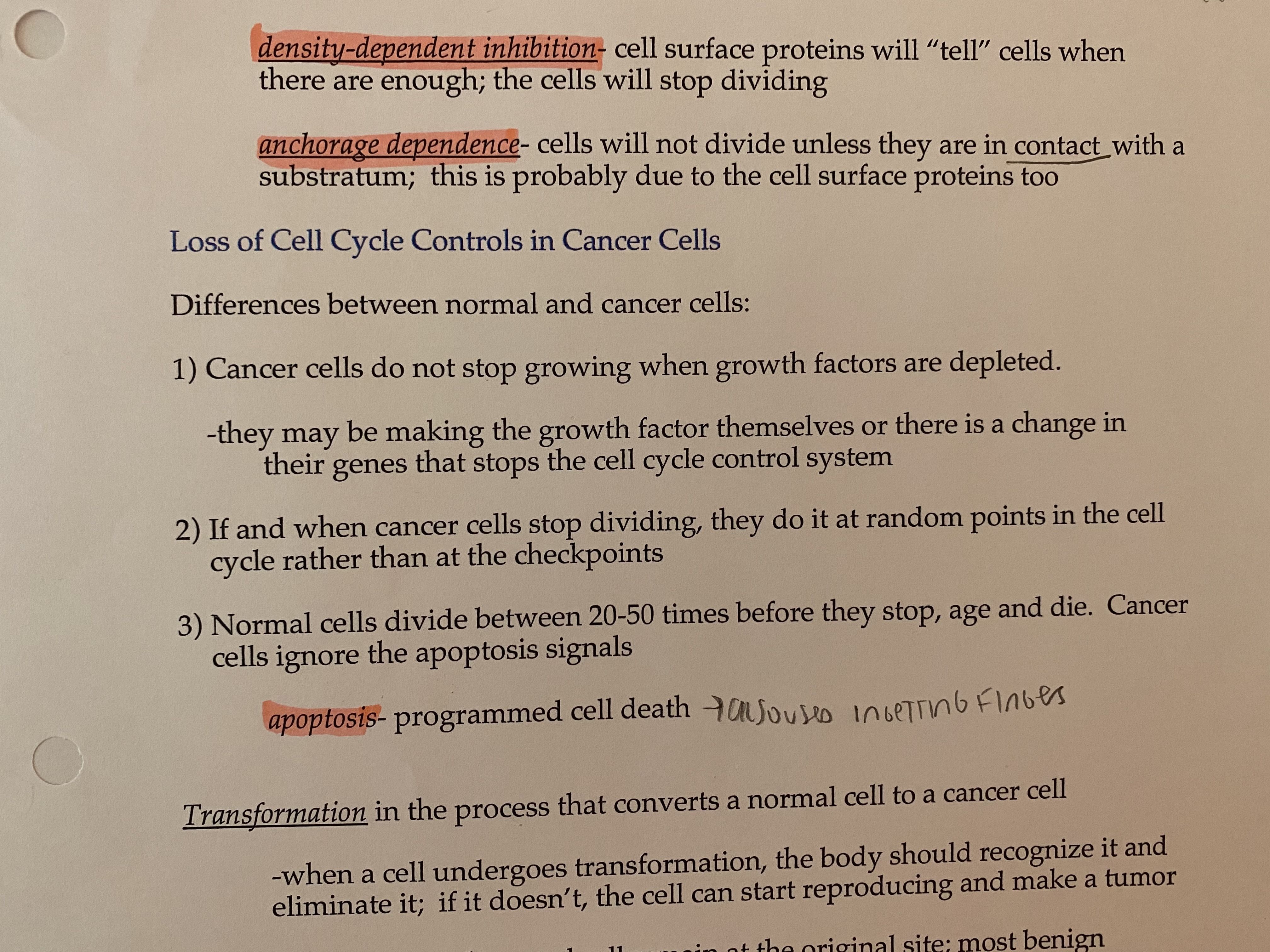
| density-dependent inhibition | cell surface proteins will "tell" cells when there are enough; the cells will stop dividing |
| anchorage dependence | cells will not divided unless they are in contact with a substratum; this is probably due to the cell surface proteins too |
| apoptosis programmed cell death - lack of this causes cancer because cells reproduce unregulated | |
| benign tumor | abnormal cells remain at original site, most benign tumors cause no problem and can be removed by surgery |
| malignant tumor | cancer cells metabolism can be changed due to chromosomal changes - since they cannot connect to neighboring cells due to changes in the chromosomal instructions, cancer cells may spread to other parts of the body, AKA metastasis - cancer cells may also secrete signaling molecules that will cause blood vessels to grow toward the tumor |
Want to create your own Flashcards for free with GoConqr? Learn more.