26683296
Description
Flashcards by Joshua Nunn, updated more than 1 year ago
|
|
Created by Joshua Nunn
about 5 years ago
|
|
| Question | Answer |
| Inferior Border of the Mandible - Appears as a heavy, broad band of bone - Radiopaque | |
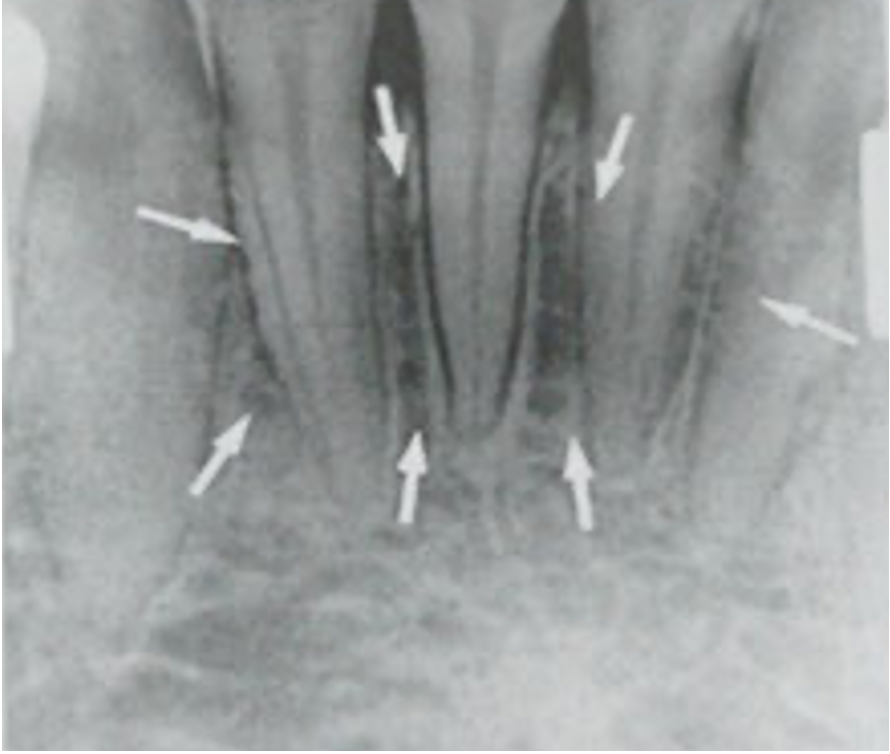
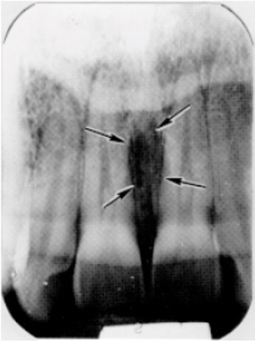
| Genial Tubercle + Lingual Foramen - Appears on lingual surface of the mandible just above the inferior border and immediately below and between the two central incisors - Genial tubercle is the radiopaque ring that surrounds the radiolucent lingual foramen | |
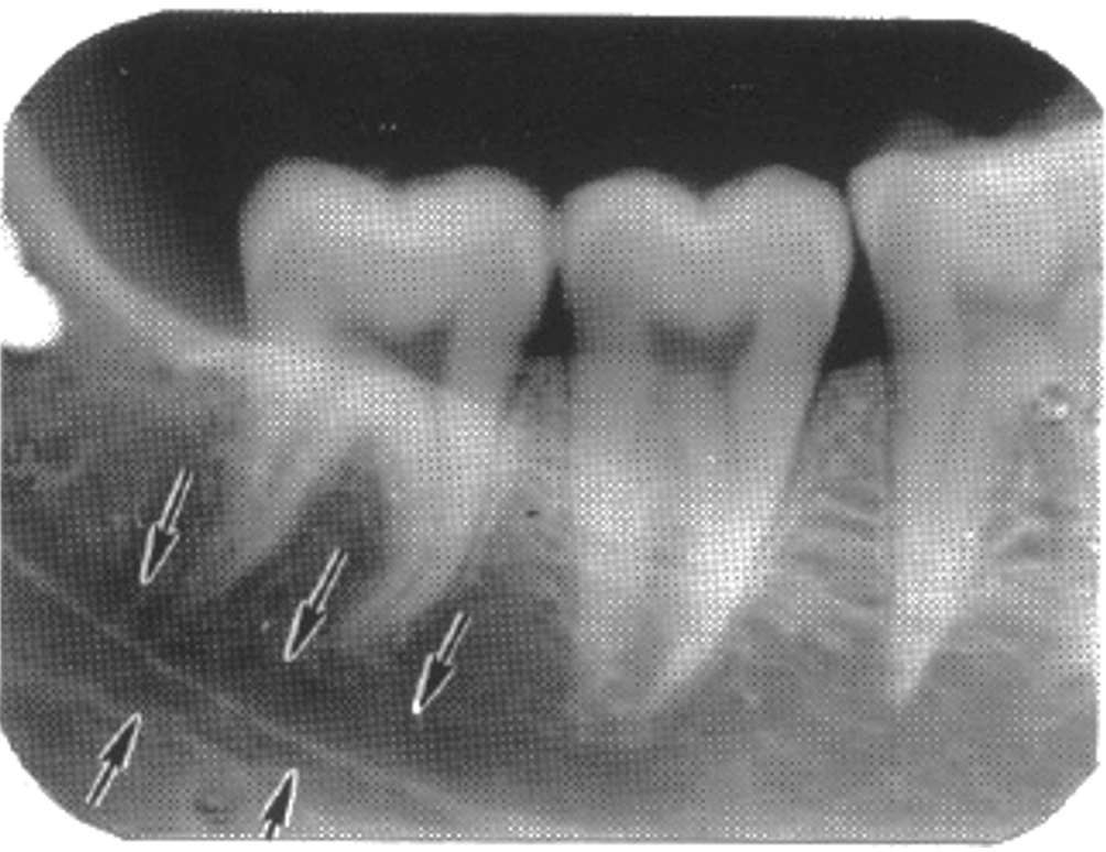
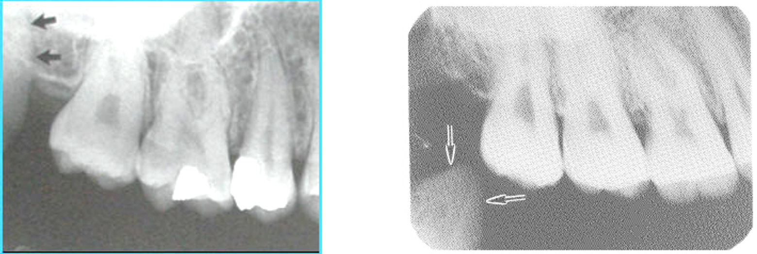
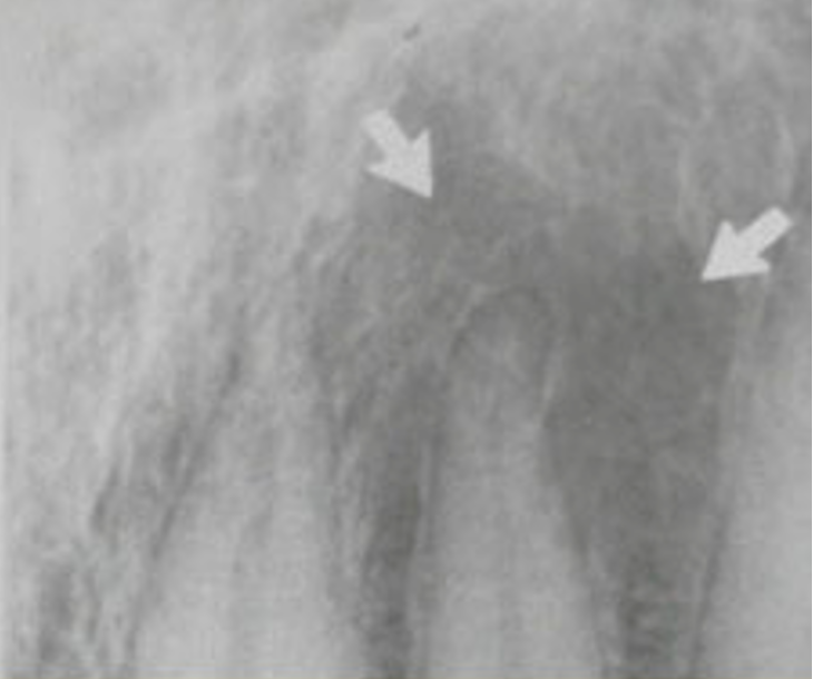
| Mental Foramen - Located below the first molar -> first premolar (varies between patients) but can be superimposed onto the tooth by poor angulation - Examine the continuity of the lamina dura to determine if it is a pathological condition or the mental foramen | |
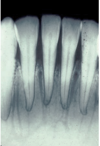
| Mental Ridge - Appears on pA's of mandibular incisors - Bilateral "V" shape located inferior to tooth apices but may be superimposed over them | |
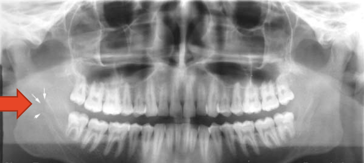
| Mandibular Foramen - Radiolucent area at the ramus of the mandible | |
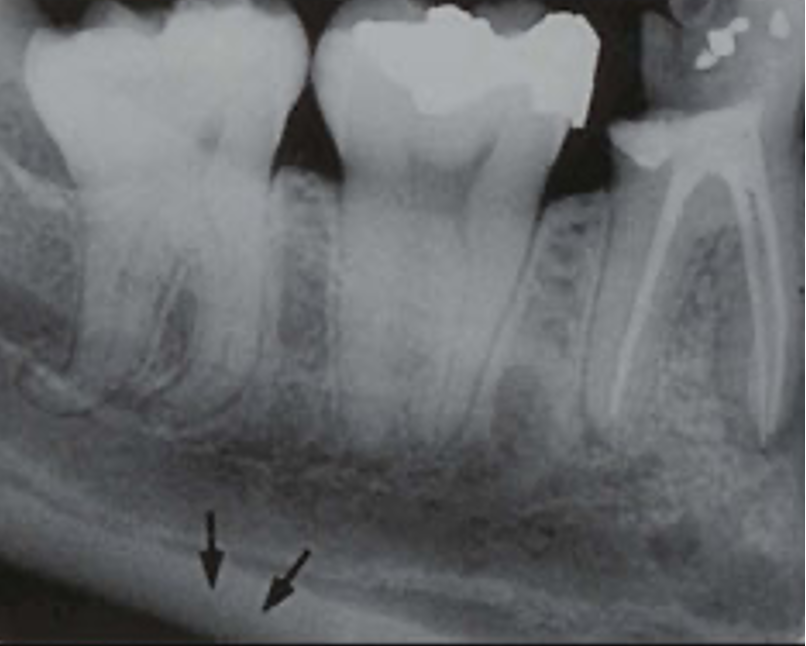
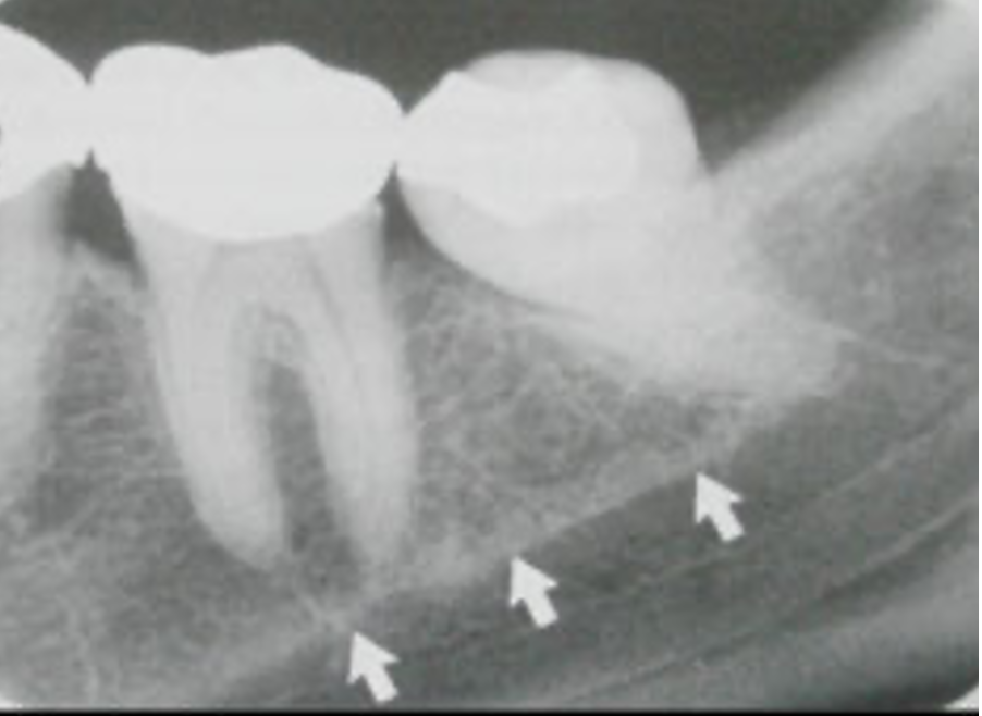
| Mandibular Canal - Begins at the mandibular foramen and terminates at the mental foramen - Appears as a radiolucency above the inferior border - Borders of the canal MAY be apparent as thin radiopaque lines | |
| Mental Fossa - Depression on the labial surface of the mandible around the midline and above the mental protuberance - Appears as a generalised diffuse radiolucency as the bone here is thinner | |
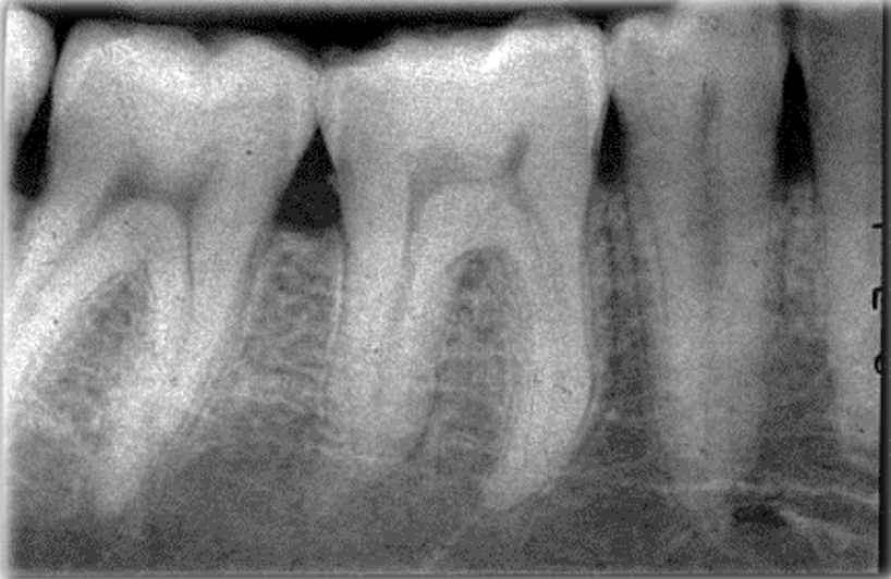
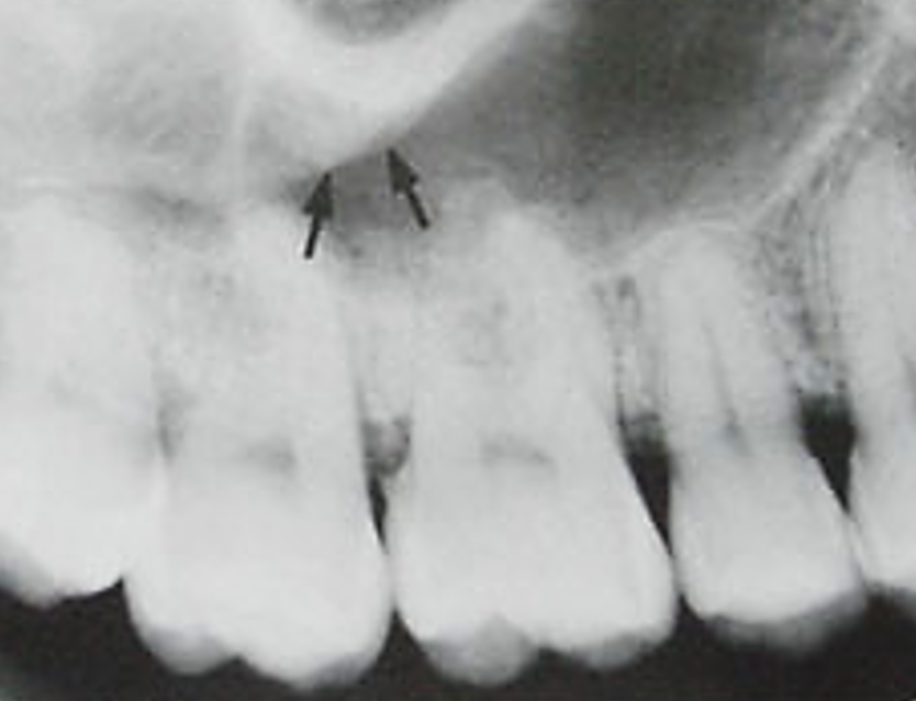
| Nutrient Canals - Not apparent in radiographs of ALL patients, but may appear in pA's of mandibular incisors - Represent the nutrient canals of the bone, appearing fairly uniform in width and running vertically between the incisors | |
| Mylohyoid Ridge - Also known as internal oblique ridge - Radiopaque line extending from the chin to the molar region towards the ramus | |
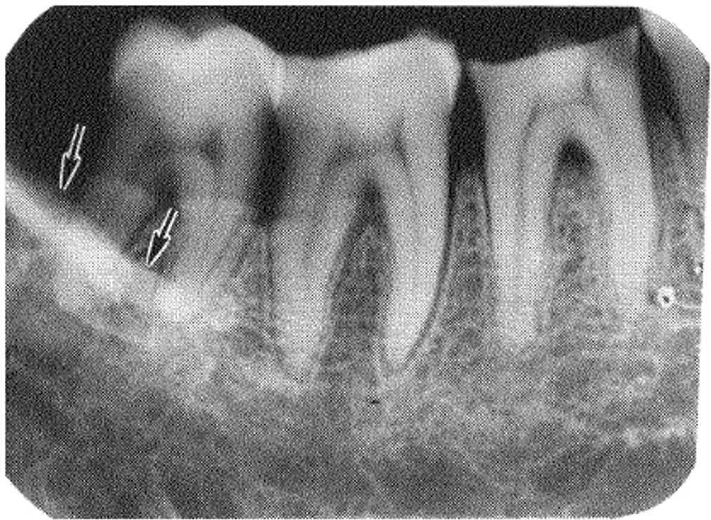
| External Oblique Ridge - Anterior portion of the descending ramus - Varying width and density (may be so dense that it obscures tooth roots | |
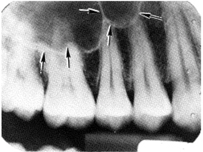
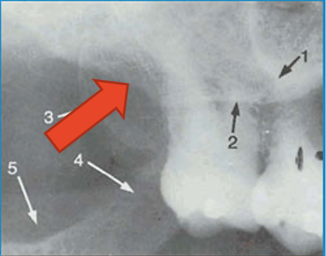
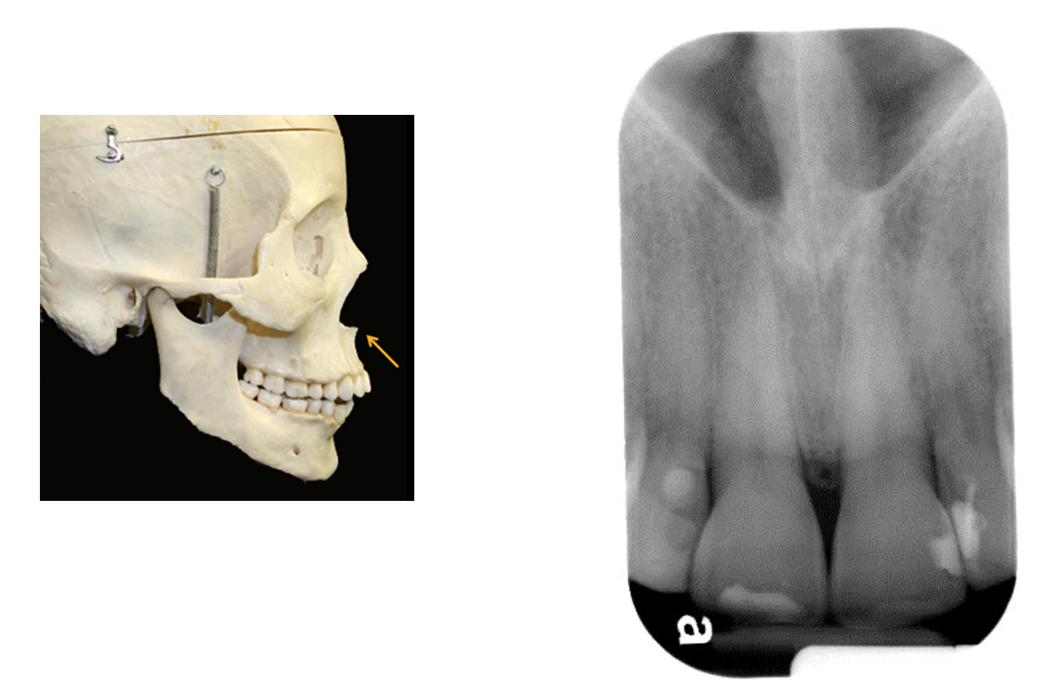
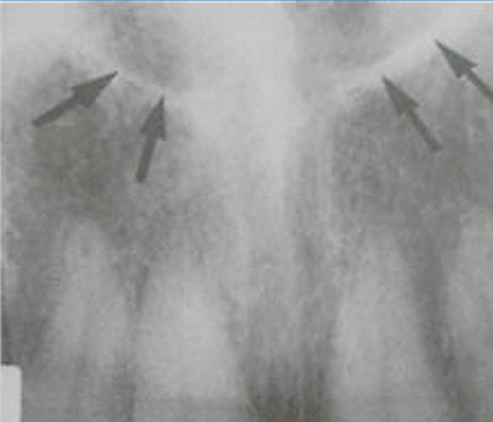
| Maxillary Sinus - Radiolucent dark area above the maxillary molars with radiopaque borders/margins - Huge variability in sizes, enlarge during childhood maturing around 15-18 - Floor can appear above the apices or well below (extremely variable) - Occasionally the roots will project into the sinus (take this into account when extracting) | |
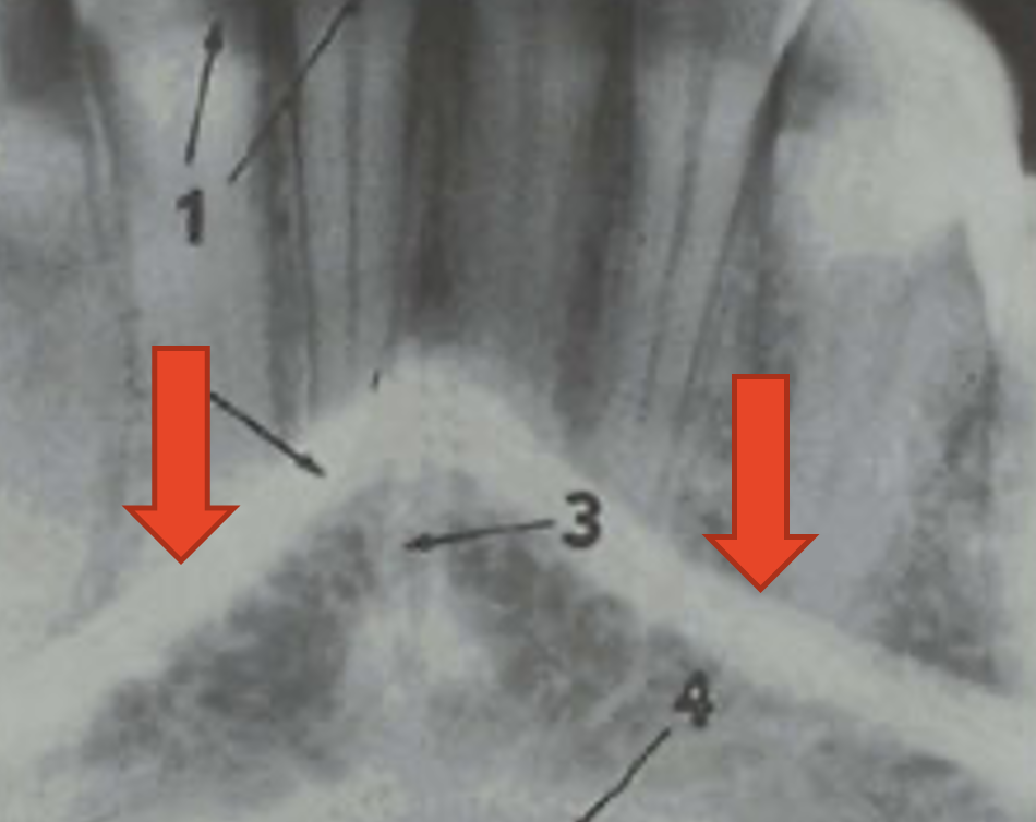
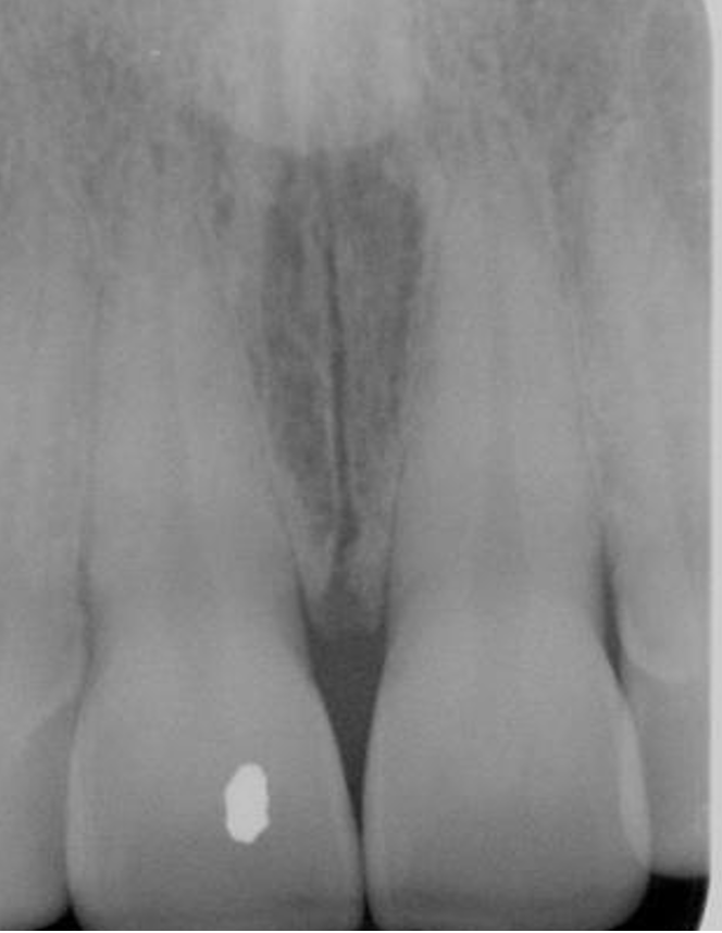
| Intermaxillary Suture - Also known as the median palatine suture - May be mistaken for a fracture - Thin and irregular radiolucent line usually starting between the 11 and 21, extending across the hard palate | |
| Nasolacrimal Canal - Round/oval radiolucent area appearing on occlusal projections of upper molars | |
| Coronoid Process of the Mandible - Triangular grey area - Can be superimposed ONTO upper teeth | |
| Zygomatic Process and Zygomatic Bone - Dense radiopacity - Apparent in pA's of upper molars, especially over the first and second molars | |
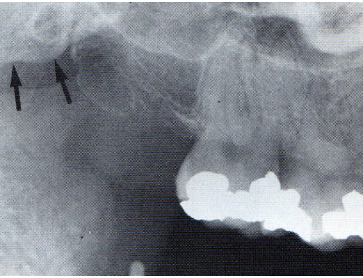
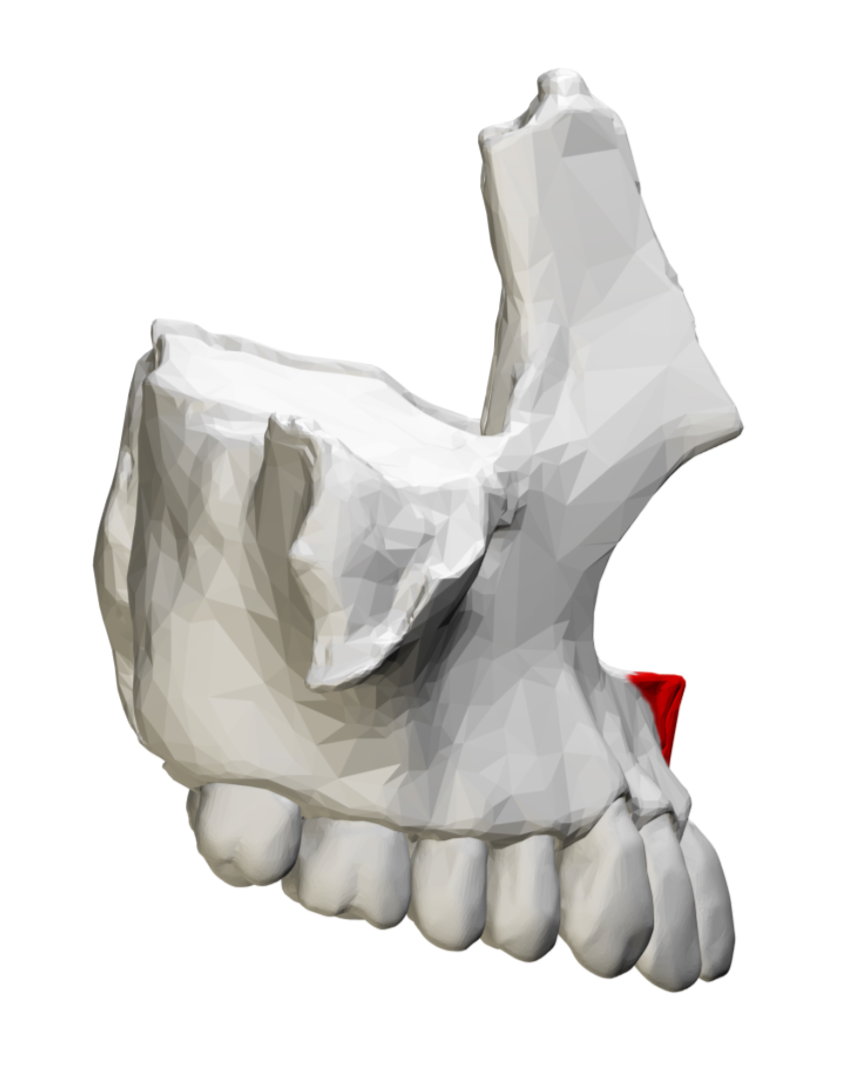
| Maxillary Tuberosity and Surrounding Structures - Appears as normal bone distal to the last molar - Curves up and distally at the end | |
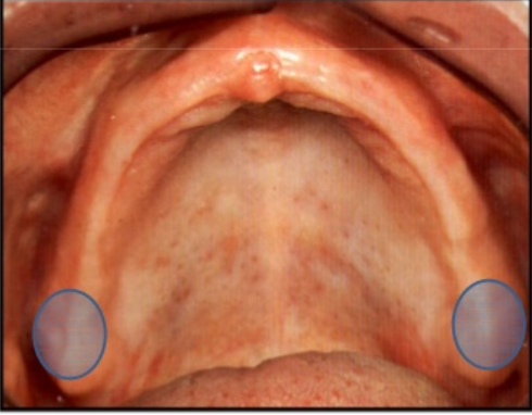
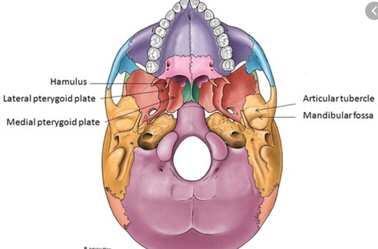
| Pterygoid Plates and Hamulus - Grey structures distal to the maxillary tuberosity on molar radiographs - Medial and lateral pterygoid plates + hamulus of the sphenoid bone | |
| Soft Tissue Shadows - Formed by the tip of the nose - Typical and frequent on central and lateral incisors | |
| Lateral Fossa - Gentle depression in the apex of the maxillary lateral incisor - Present in pA's as an ill-defined radiolucency | |
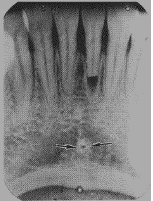
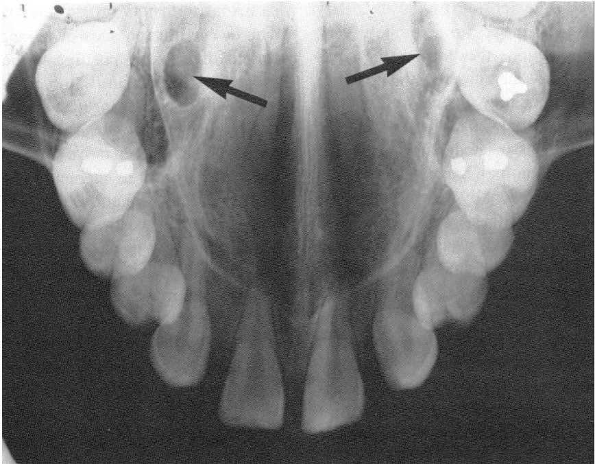
| Incisive Foramen - Usually oval shaped but varies greatly - Found between the central incisors, located on the hard palate - Appears in radiographs on the midline, between the two roots of the central incisors | |
| Anterior Nasal Spine and Nasal Septum - Anterior nasal spine appears as a "V" in the midline | |
| Floor of the Nasal Cavity - Appears on radiographs above the anterior nasal spine |
Want to create your own Flashcards for free with GoConqr? Learn more.